Microbiology Laboratory Turkey

Mikrobiyoloji Ile Ilgili Tüm Konuların Kısa ve Öz Anlatımları. Microbiology Lab Information.

Bu Blogda Ara(SEARCH)

MicroLab

parasites egg etiketine sahip kayıtlar gösteriliyor. Tüm kayıtları göster

9 Mart 2019 Cumartesi

Mart 09, 2019

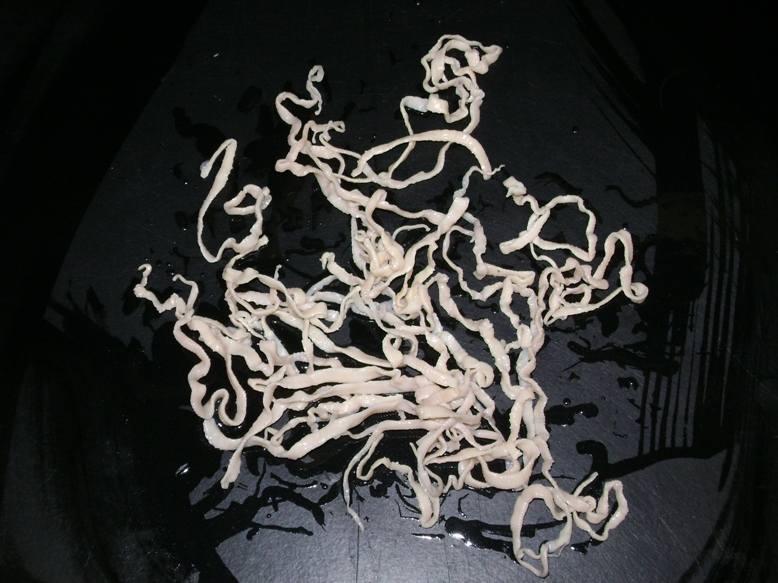
HYMENOLEPIS DIMINUTA (RAT TAPEWORM)

HYMENOLEPIS DIMINUTA
(RAT TAPEWORM)

Hymenolepis diminuta differs from Hymenolepis nana in that: 
♦ The adult worm measures about 10-60 cm 
♦ The rosetellum on the head has no hooks 
♦ In the mature segment, there are two testes at one side and another testis on the other side.
Life Cycle
The adult worms are present in the small intestine of man and rats. Eggs passed in stool are similar to the eggs of H. nana but are brown in color with no polar filaments arising from the polar thickening. The eggs are ingested by the rat flea where they develop to cysticercoid stage. Infection to man takes place accidentally by food or contaminated hands by cysticercoid stage.
Pathogenecity
Most infections are asymptomatic, but occasionally, patients may present with nausea, anorexia and diarrhea.

Treatment
Same as Hymenolepis nana.

INSTAGRAM

22 Şubat 2019 Cuma

Şubat 22, 2019

Necator americanus

 Necator americanus

This species, so called American hookworm, is found in predominantly the tropics. The anterior end is hooked against the body curvature. The mouth is provided ventrally and dorsally with cutting plate.


Morphology
Male:
The male measures 8 cm in length. The posterior end is broadened into a membraneous copulatory bursa, which is provided with two long spicules fused distally.  

Female:
The female measures 10 cm in length. The posterior end is straight  
Infective stage and methods of infection: 
The filariform larva infects by skin penetration.


Life Cycle
Adult male and female worms live in the small intestine. The female lays eggs (oval, 60x40 microns), which contain immature embryo in the 4 cell stage. When the eggs pass in the stool to the soil and under favorable conditions of temperature, moisture and oxygen, they hatch into larvae, which molt twice and become infective. When the filariform larvae penetrate the skin, they circulate in the blood, reach the lungs, ascend to the trachea, descend to esophagus to reach the small intestine and become adults.


Pathogenecity
Adult worms in the intestine feed on blood causing iron deficiency anemia. The larvae may cause inflammation of the lungs. 
Diagnosis: 
Examination of stool by direct saline smear to detect the eggs.


INSTAGRAM

18 Şubat 2019 Pazartesi

Şubat 18, 2019

TAENIA SOLIUM (PORK TAPEWORM)

TAENIA SOLIUM


 The adult worms of T. solium reside or inhabit the upper jejunum. Infection has worldwide distribution.  


Morphology: 
Adult worm measures about 3 meters in length. The globular scolex has rostellum with 2 rows of hooklets. There are <1000 proglottids. Gravid proglottid liberates about 30,000-50,000 eggs. 
Life Cycle 
Embryonated eggs passed with stool are ingested by pig and the embryo is released. It penetrates the intestinal wall and is carried by vascular channels to all parts of the body. After a period of 2-3 months of development the encysted larval stage called cysticerci or bladder worm occurs in the striated muscles of the tongue, neck, trunk brain, eye, and the nervous system. The cysticercus survives for 5 years. Humans become infected by eating pork containing larvae, cysticercus cellulosae. When improperly cooked cysticercus infected meat is eaten by man, the scolex remains undigested and attaches itself to the intestinal wall and chain of proglottids begin to grow to adult worm.

Clinical Manifestations 
Resembles that of T. saginata infection.
Diagnosis 
Demonstration of eggs in stool specimen.


Prevention: 
♦ Treatment of infected persons. 
♦ Thorough cooking of pork and proper processing 
♦ Proper disposal of human excreta (good hygiene/sanitation).


INSTAGRAM

14 Şubat 2019 Perşembe

Şubat 14, 2019

Ancylostoma duodenale

Ancylostoma duodenale

Grayish-white in color. The body is slightly ventrally curved. The anterior end follows the body curvature. The buccal cavity is provided ventrally with pairs of teeth and dorsally with a notched dental plate.  Distribution: This species is found in the northern part of the world including China, Japan, Europe, North Africa and Ethiopia.

Morphology 

Male: 
The male measures 10 cm in length. The posterior end is broadened into a membraneous copulatory bursa that is provided with two long spicules. 

Female: 
The female measures 12 cm in length. The posterior end is straight. 


INSTAGRAM