Microbiology Laboratory Turkey

Mikrobiyoloji Ile Ilgili Tüm Konuların Kısa ve Öz Anlatımları. Microbiology Lab Information.

Bu Blogda Ara(SEARCH)

MicroLab

Escherichia coli etiketine sahip kayıtlar gösteriliyor. Tüm kayıtları göster

25 Şubat 2019 Pazartesi

Şubat 25, 2019

Membrane Filter Technique | Membrane Filtration Method (Microbes)

Membrane Filter Technique | Membrane Filtration Method

Microbes are considered as an important microorganisms, its have different types, sizes and uses, Bacteria for example can be found everywhere; it has sizes from 0.2 µm to around 3 µm. Coliform bacteria testing known as the most microbes detected for water, and the presence of Escherichia coli (size: 1 µm) is an indication for the presence of pathogens that leads to water contamination. Membrane Filtration Technique is the popular method for the detection of many microbes, it can detect up to 300 Colony Forming Unit (CFU), and it has the ability to detect different types according to media and sample types.

Membrane Filtration need the following equipment in general for work:
Laminar Flow Hood (Optional).
Vacuum Pump with Rubber Tube.
Suction Flask 1 L.
Nitrocellulose Filter, Pore Size: 0.45 µm.
Microbiology Media.
Sterile Distilled Water.
70% Ethanol.
Flame.
Forceps.
Incubator.
Autoclave.
Tissue Paper.
Sterile cups or Sterile bags.
Suction Funnel.

Sample Collection:
Sample types and collection methods are different, Samples may be water, beverage, swab, sugar, or syrup. In this article an explanation of water sample will done. Water samples are collected from tanks as follow:
Wet piece of tissue paper by 70% ethanol and wipe the valve of the tank.
Open the valve to let some water out to erase any excess of ethanol then close it.
Open the sterile bag or sterile cup in the same time open the valve and take 100 ml then close the valve and the bag / cup.
Transfer the sample to Microbiology lab immediately.
Water Samples can be stored in refrigerator at 4°C to inhibit the growth and the rise in microbial numbers.

Working Procedures:
    Disinfect Laminar flow hood and clean all equipment using 70% ethanol.
    Turn on UV lamp and take time according to laminar hood type.
    Turn on air flow (that will turn off UV lamp), then clean all the tools again by 70% ethanol.
    Enter water sample inside the hood and clean it by 70% ethanol.
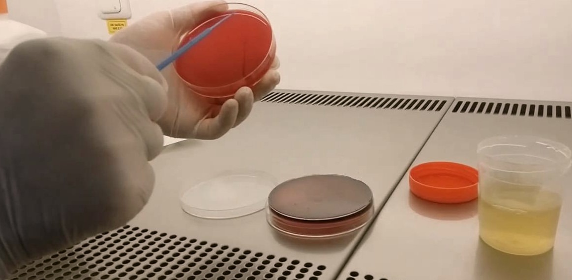
    Put the filter on the suction funnel and pour half of the sample then turn on the pump and pour the other half.
    Turn off the pump when all sample content filtered.
    Mount the filter on media plate and incubate it on the proper temperature inside the incubator.


    19 Şubat 2019 Salı

    Şubat 19, 2019

    Aglütinasyon Testi

    Aglütinasyon Testi

    Kültürlerde, bir bakterinin Salmonella veya Shigella türlerinden biri olarak karar verilmesi için mutlaka lam aglütinasyonu yapılması gerekir. Ayrıca, Escherichia coli’nin serotipleri de (Serotip, özellikle bakteri ve virüslerde, antijen karakterleri ile belirlenen türün alt tür kümesidir.) ancak, lam aglütinasyonu yapılarak tespit edilir. Lam aglütinasyonu aynı zamanda bir serolojik doğrulama testidir. 

    Testin Yapılış Tekniği; 
    • Öncelikle bakteri süspansiyonu hazırlanır; bir tüpe konulan birkaç damla tuzlu suya, katı besiyerinde oluşmuş kolonilerden özeyle birkaç tane alınıp ezilerek süt görünümünde karışım oluşturulur. 
    • Kullanılacak antiserumlar, 1/5 ya da 1/10 oranında sulandırılarak kullanılır. (Antiserum; enfeksiyon yapıcı mikroorganizmalara ya da zehirli maddelere karşı etkili özgül antikorları içeren kan serumudur.)
    • Temiz bir lam alınır. Pastör pipetiyle lamın bir ucuna bir damla serum fizyolojik, diğer ucuna bir damla antiserum damlatılır. 
    • Her iki damla üzerine, ince bir pastör pipetiyle bakteri süspansiyonundan birer damla damlatılır. 
    • Lam elle tutulup eğdirilerek bir dakika beklenir.
    • Sonuç, siyah zemin üzerine tutularak değerlendirilir. Kümeleşerek parçacıkların oluşması, aglütinasyonun olumlu sonuçlandığını gösterir.

    31 Ocak 2019 Perşembe

    Ocak 31, 2019

    Gram Negative Bacteria

    Gram Negative Bacteria

    Gram negative bacteria are bacteria that do not retain the crystal violet dye in the Gram stain protocol. Gram negative bacteria will thus appear red or pink following a Gram stain procedure due to the effects of the counterstain (for example safranin).

    The Gram Stain

    In microbiology, the visualization of bacteria at the microscopic level is facilitated by the use of stains, which react with components present in some cells but not others. This technique is used to classify bacteria as either Gram-positive or Gram-negative depending on their colour following a specific staining procedure originally developed by Hans Christian Gram. Gram-positive bacteria appear dark blue or violet due to the crystal violet stain following the Gram stain procedure; Gram-negative bacteria, which cannot retain the crystal violet stain, appear red or pink due to the counterstain (usually safranin).


    The reason bacteria are either Gram-positive or Gram-negative is due to the structure of their cell envelope. (The cell envelope is defined as the cell membrane and cell wall plus an outer membrane, if one is present.) Gram-positive bacteria, for example, retain the crystal violet due to the amount of peptidoglycan in the cell wall. It can be said therefore that the Gram-stain procedure separates bacteria into two broad categories based on structural differences in the cell envelope.

    Cell Envelope of Gram Negative Bacteria

    The Gram negative cell envelope contains an additional outer membrane composed by phospholipids and lipopolysaccharides which face the external environment. The highly charged nature of lipopolysaccharides confer an overall negative charge to the Gram negative cell wall. The chemical structure of the outer membrane lipopolysaccharides is often unique to specific bacterial strains (i.e. sub-species) and is responsible for many of the antigenic properties of these strains. Many species of Gram-negative bacteria are pathogenic. This pathogenicity is often associated with the lipopolysaccharide (LPS) layer of the Gram-negative cell envelope.

    Characteristics of Gram Negative Bacteria

    Gram-negative bacteria have a characteristic cell envelope structure very different from Gram-positive bacteria. Gram-negative bacteria have a cytoplasmic membrane, a thin peptidoglycan layer, and an outer membrane containing lipopolysaccharide. There is a space between the cytoplasmic membrane and the outer membrane called the periplasmic space or periplasm. The periplasmic space contains the loose network of peptidoglycan chains referred to as the peptidoglycan layer.

    Gram Negative Bacteria
    • Acinetobacter
    • Actinobacillus
    • Bordetella
    • Brucella
    • Campylobacter
    • Cyanobacteria
    • Enterobacter
    • Erwinia
    • Escherichia coli
    • Franciscella
    • Helicobacter
    • Hemophilus
    • Klebsiella
    • Legionella
    • Moraxella
    • Neisseria
    • Pasteurella
    • Proteus
    • Pseudomonas
    • Salmonella
    • Serratia
    • Shigella
    • Treponema
    • Vibrio
    • Yersinia


    INSTAGRAM

    4 Ocak 2019 Cuma

    Ocak 04, 2019

    Urinary Tract Infection (UTI)

    Urinary Tract Infection (UTI)


    Reddening caused by urinary tract infection of the urinary tract lining, and consequently cause irritation and pain, urinary disturbance and frequent blood is causing symptoms such as the need to urinate. If the infection spreads to your kidneys can become a serious health problem.

    The urinary tract is one of the body's fluid waste products which produce and store urine. Normal urine bacteria (germ) is not.However, sometimes the bacteria enters the urinary tract outside the body and causes infection and inflammation. This is a urinary tract infection. Infection urethra (urethritis called state), kidneys (pyelonephritis name given situation) or bladder (a condition called cystitis) as appropriate. Cystitis is the most common type of urinary tract infection.

    WHAT ARE THE SYMPTOMS OF URINARY INFECTION?
    A urinary tract infection, reddening of the lining of the urinary tract, causing irritation, which may form part of the following symptoms:
    - Side (abdomen), abdomen or pain in the pelvic region 
    - Sub-pelvic pressure 
    - the need for Frequent Urination 
    - painful urination (dysuria) 
    - Urine emergency exit needs 
    - Incontinence (urine leakage) 
    - Night of the need for frequent urination 
    - Abnormal urine color 
    - Blood in the urine 
    - Strong or foul-smelling urine


    Other Symptoms That Can Be Associated With Urinary Tract Infections Include:

    - Pain in the side of the body 
    - Fatigue 
    - Fever 
    - Shivering 
    - Vomiting 
    - Mental confusion


    CAUSES Urinary Tract Infection?

    Urinary tract infections, urinary tract and bladder that causes inflammation and entering the microorganisms (usually bacteria ) to occur.

    More than 90 percent of cases cystitis, normally in the gut bacteria is from E.coli caused.


    HOW Urinary Tract Diagnosed?
    A urinary tract infection following tests are used to diagnose:
    Urinalysis:  Red blood cells, white blood cells and examines urine for bacteria (number of white and red blood cells indicates an infection.)
    Urine culture to determine the type of bacteria in the urine :  This is important to help determine the appropriate treatment.

    Infection does not respond to treatment or if you have recurring infections, urinary be done in terms of the following tests to examine your way through disease or injury:


    Intravenous Pyelogram (IVP):  After a special dye bladder, kidney, ureter and an array of X-ram is injected. (paint, helps you look better on X-ray).
    Ultrasound:  A test that uses sound waves to create images of internal organs
    Cystoscopy:  The urethra through a lens and a light source to see inside the bladder (cystoscopy) equipped with a test that uses a special tool.
    CT Scan:  Body sections (such as strips) an X-ray field type (the typical X-ray is more sensitive)

    Urinary Tract HOW IS IT TREATED?
    Antibiotics are drugs that kill bacteria, used to treat urinary tract infections. Your doctor will choose the bacteria that cause you to enfeksiyonunları a treatment drug. Antibiotics are commonly used include:
    - Nitrofurantoin (A preparation antibiotics that fight bacteria in the body) 
    - sulfonamides (sulfa drugs - antibacterial are composed of sulfonic acid) 
    - Amoxicillin (causes of death by disrupting the cell walls of bacteria) 
    - Cephalosporins (both structural and antibiotics resembling penicillin its effects both) 
    - Trimethoprim / sulfamethoxazole 
    - Doxycycline (usually recommended for treatment of acne and infectious) 
    - Quinolones (obtained from viable microorganisms that many antibiotic is obtained by chemical means as opposed to synthetic substances)
    Adhere strictly to the instructions of your doctor while taking medication is very important. Antibiotics do not leave prematurely. If the infection is completely cured with antibiotic treatment, the risk of recurrence is high.