üre testi
Eylül 30, 2018
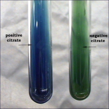
ÜRE TESTİ (UREASE TEST)
ÜRE TESTİ (UREASE TEST)
Bu test, bakterilerin üreyi hidrolize eden üreaz (urease) enzimini belirlemek
amacıyla yapılır. Bu bakteriler, üreaz enzim aktiviteleri ile üreyi parçalayarak karbondioksit
ve amonyak oluşturur. Oluşan alkali ortamda, besiyerinin pembeye dönmesine neden olur.
Testte Christensen’in üre içeren besiyerleri (üreli buyyon veya agar) kullanılır.
Proteus
mirabilis ve Klebsiella üreaz pozitif Escherichia coli negatif bakterilerdir.

Testin yapılış tekniği;
➨ Mikroorganizma kültürlerinden, petri kutusu veya tüpteki Christensen’in üreli
agar yüzeyine tekniğine uygun ekim yapılır.
➨ Etüvde, 37°C’de 1-5 gün inkübe edilir.
➨ Renk değişmelerine dikkat edilerek her gün kontrol edilir. (Bazı durumlarda
renk değişikliği 5-6 saat içinde meydana gelebilir.)
➨ Kolonilerde veya tüpte pembe rengin meydana gelmesi pozitif reaksiyonu
gösterir.
Negatif durumda renk değişikliği olmaz.

Üre Testi Pozitif Bakteriler
- Proteus spp
- Corynebacterium spp
- Helicobacter pylori
- Brucella spp

INSTAGRAM
FACEBOOK