Salmonella is a Gram-negative facultative rod-shaped bacterium in the same proteobacterial family as Escherichia coli, the family Enterobacteriaceae, trivially known as "enteric" bacteria. Salmonella is nearly as well-studied as E. coli from a structural, biochemical and molecular point of view, and as poorly understood as E.coli from an ecological point of view. Salmonellae live in the intestinal tracts of warm and cold blooded animals. Some species are ubiquitous. Other species are specifically adapted to a particular host. In humans, Salmonella are the cause of two diseases called salmonellosis: enteric fever (typhoid), resulting from bacterial invasion of the bloodstream, and acute gastroenteritis, resulting from a foodborne infection/intoxication.
SALMONELLA on XLD AGAR
Diagnosis
Salmonellosis should be considered in any acute diarrheal or febrile illness without obvious cause. The diagnosis is confirmed by isolating the organisms from clinical specimens (stool or blood).
Diagnosis
The diagnosis of salmonellosis requires bacteriologic isolation of the organisms from appropriate clinical specimens. Laboratory identification of the genus Salmonella is done by biochemical tests; the serologic type is confirmed by serologic testing. Feces, blood, or other specimens should be plated on several nonselective and selective agar media (blood, MacConkey, eosin-methylene blue, bismuth sulfite, Salmonella-Shigella, and brilliant green agars) as well as intoenrichment broth such as selenite or tetrathionate. Any growth in enrichment broth is subsequently subcultured onto the various agars. The biochemical reactions of suspicious colonies are then determined ontriple sugar iron agar and lysine-iron agar, and a presumptive identification is made. Biochemical identification of salmonellae has been simplified by systems that permit the rapid testing of 10–20 different biochemical parameters simultaneously. The presumptive biochemical identification of Salmonella then can be confirmed by antigenic analysis of O and H antigens using polyvalent and specific antisera. Fortunately, approximately 95% of all clinical isolates can be identified with the available group A-E typing antisera. Salmonella isolates then should be sent to a central or reference laboratory for more comprehensive serologic testing and confirmation.
Klebsiella; toprakta, suda, insan ve hayvanların bağırsağında, üst solunum yollarında bulunur. Bu grupta; Klebsiella pneumoniae, Klebsiella rhinosclecomatis, Klebsiella ozanae gibi bakteriler vardır.
Morfolojisi
Klebsiella pneumoniae hareketsiz, sporsuz, geniş kapsüllü, gram negatif (-) basildir. Genellikle uç uca, ikişer ikişer kısa zincirler halinde ve tek tek bulunur. Bakteriyolojik boyalarla iyi boyanır.
Üreme Özellikleri
Klebsiella pneumoniae, aerob ve fakültatif anaerobdur. Optimal üreme ısısı 37oC, optimal pH‟sı 7.4‟tür. Kuruluğa dirençli olup özellikle organik maddeler içinde kurutulurlarsa aylarca canlı kalır. Isıya dayanıksız olup nemli ısıda 55 oC‟de yarım saate ölür. Sıvı besiyerinde homojen bir bulanıklık, yüzeyde zar ve dipte mukoid bir çöküntü oluşturarak ürer. Katı besiyerinde mukuslu ve kapsüllü olduğu için öze ile alındığında sünen M tipi koloniler oluşturur; kanlı jelözde hemoliz oluşturmaz. İndol, H2S, Voges-Proskauer, metil kırmızısı testleri negatif,üre ve KCN testleri pozitif olan bakterilerdir.
Yaptığı Hastalıklar
Klebsiella pneumoniae; bulunduğu yerlerde uygun koşulların oluşması veya yer değiştirerek diğer organ ve sistemlere yerlemeleri halinde, başta pnömoni olmak üzere, anjin, sinüzit, otitis media, üst solunum yolu enfeksiyonu, gastroenterit, septesemi, menenjit, kolesistit ve üriner sistem hastalıklarına neden olur.
Laboratuvar Teşhisi ➨ İnceleme örnekleri: İdrar, balgam, kan, dışkı, boğaz sürüntüsü, cerahat. ➨Teşhis yöntemleri
➧ Mikrobiyolojik örneğin direkt mikroskobik incelemesi; santrifüj edilen idrardan hazırlanan preparatın incelenmesinde; bol lökosit ve ikişerli hareketsiz bakterilerin görülmesiyle ve hazırlanan preparatın gram boyasıyla boyanarak incelenmesiyle de teşhis edilir.
➧ Kültür;EMB AGAR, Endo, Mac-Conkey Agar besiyerine ekim yapılır.
➧ Biyokimyasal Testler; tipik mukoid kolonilerden çeşitli biyokimyasal testlerle teşhis edilir.
Amaç, B grubu Streptococcusların oluşturduğu CAMP (Christie, Atkins, Munch-Peterson) faktörünü tespit etmektir. Bu testi bulan bilim adamlarının ilk harfleri alınarak CAMP adı oluşturulmuştur.
Testin yapılış Tekniği;
➤ Bazı Staphylococcusların (Staphylococcus aureus) var olduğu düşünülen örnekten,
özeyle alınarak %5‟lik koyun kanlı agarın ortasına boydan boya kalın bir çizgi
şeklinde ekilir.
➤ Bu ekim çizgilerine dik doğrultuda, Staphylococcus ekim çizgisine dokundurmamaya
dikkat ederek incelenecek mikroorganizmadan 2-3cm uzunlukta ekim yapılır.
➤ Plaklar etüvde, 37°C'de 18-24 saat inkübe edilir.
➤ İnkübasyon sonunda, Streptococcus ve Staphylococcusların üreme yapıları incelenir.
Birbirlerine yakın oldukları yerde ok başı şeklinde hemoliz meydana gelirse
CAMP testi pozitif olarak değerlendirilir.
CAMP testinde ok başı şeklindeki görünen pozitif sonuç
The coloration of bacteria by applying a single staining
solution to a fixed smear is a “simple staining”. It is
called as “monochrome staining” because only one dye
is used. Basic dyes like methylene blue, crystal violet,
safranine, malachite green are used to determine the
shape, size and arrangement of bacteria.
PROCEDURE:
➤ Prepare the smear and heats fix it.
➤Flood the smear with methylene blue stain and allow it
to react for 1 minute.
➤Wash the slide with distilled water, blot and air dry.
To determine the ability of the organism to hydrolyse urea by the action of urease enzyme.
Principle
Urea is a nitrogen-containing compound that is produced during decarboxylation of the amino acid arginine in the urea cycle. Urea is a major organic waste product of protein digestion in most vertebrate and is excreted in urine. Some bacteria have the ability to produce an enzyme urease as part of its metabolism to break down urea. The urease is a hydrolytic enzyme which attacks the carbon and nitrogen bond with the liberation of ammonia and carbon dioxide. It is a useful diagnostic test for identifying bacteria, especially to distinguish members of the genus Proteus from Gram-negative pathogens. Proteus vulgaris is an important and fast producer of urease.
Urease test is performed by growing test organism on urea agar slant or urea broth with phenol red as an indicator with pH6.8. During the incubation period, the organism capable of producing urease enzyme hydrolyse urea and produce ammonia that raises the pH level. As the pH increases, the phenol red changes from yellowish orange as the initial colour of medium to deep pink. Failure of development of a pink colouration due to no ammonia production is evidence of an inability of the organism to produce urease enzyme.
Media
Enzymatic digest of gelatin (1 g), dextrose (1 g), NaCl (5 g), KH2PO4 (2 g), urea (20 g), phenol red (0.012 g), per 1000 mL, pH 6.8.
Procedure
Streak the surface of a urea agar slant with a portion of a well-isolated colony or inoculate test organism on urea broth containing phenol red as the indicator.
Incubate the urea agar slant or urea broth at 37°C for 24-48 hours.
Examine the development of pink colour.
In case of unknown result incubate the tube for 7 days to check for slow urease production.
Results
Positive: Deep pink colouration, Light Orange, Magenta
Negative: No Color change, Yellowish orange colouration
Limitations
Some organisms rapidly split urea (Proteus spp, H. pylori), while others react slowly.
Urea Agar should not be used to determine the quantitative rate of urease activity, as organisms vary in their capability and rate of hydrolysis.
Prolonged incubation may cause an alkaline reaction in the medium and give the false positive result.
Bu test, bakterilerin üreyi hidrolize eden üreaz (urease) enzimini belirlemek
amacıyla yapılır. Bu bakteriler, üreaz enzim aktiviteleri ile üreyi parçalayarak karbondioksit
ve amonyak oluşturur. Oluşan alkali ortamda, besiyerinin pembeyedönmesine neden olur.
Testte Christensen’in üre içeren besiyerleri (üreli buyyon veya agar) kullanılır.
Proteus
mirabilis ve Klebsiella üreaz pozitif Escherichia coli negatif bakterilerdir.
Testin yapılış tekniği;
➨ Mikroorganizma kültürlerinden, petri kutusu veya tüpteki Christensen’in üreli
agar yüzeyine tekniğine uygun ekim yapılır.
➨ Etüvde, 37°C’de 1-5 gün inkübe edilir.
➨ Renk değişmelerine dikkat edilerek her gün kontrol edilir. (Bazı durumlarda
renk değişikliği 5-6 saat içinde meydana gelebilir.)
➨ Kolonilerde veya tüpte pembe rengin meydana gelmesi pozitif reaksiyonu
gösterir.
(Indole Test, Methyl Red Test, Voges Proskauer Test and Citrate Test)
The IMViC Tests
The IMViC tests are used to differentiate the enterics (Family Enterobacteriaceae). These are the Indole test (tryptone broth), the Methyl Red and Voges-Proskauer tests (MRVP broth) and the Citrate test (Citrate agar slants). For these IMViC tests use the enterics E.coliand Enterobacter.
The significance of these tests is that when testing drinking water for the presence of the sewage indicatorE. coli, one must be able to rule out Enterobacter aerogenes.E. aerogenesis not always associated with sewage, and its presence in water would not necessarily indicate sewage contamination.
Indole Test (Tryptone broth)
Procedure: Inoculate a loopful of bacteria into a tryptone broth. Incubate 48 hours.
Description: Tryptophan hydrolysis -Some bacteria split tryptophan into indole and pyruvic acid using the hydrolase called tryptophanase. Indole can be detected with Kovac's reagent (Indole reagent). This test is very important in differentiating E. coli(indole positive) from some closely related enteric bacteria. It also differentiatesProteus mirabilis(indole negative) from all otherProteusspecies (indole positive). Tryptone broth is used for this test as it contains a large amount of tryptophan.
Interpretation: After incubation: The broth must be turbid. A clear broth indicates that your organism did not grow and cannot be tested. Add a few drops of Indole reagent to the broth culture (tryptone broth). DO NOT SHAKE THE TUBE. A positive result has a red layer at the top. A negative result has a yellow or brown layer.
Methyl Red Test (MRVP broth) Procedure: Inoculate a loopful of bacteria into MRVP broth. Incubate 3 to 5 days.
Description: Mixed acid fermentation -Many gram-negative intestinal bacteria can be differentiated based on the products produced when they ferment the glucose in MR-VP medium. Escherichia, Salmonella, andProteusferment glucose to produce lactic, acetic, succinic, and formic acids and CO2, H2, and ethanol. The large amounts of acids produced lowers the pH of the medium -Methyl red (a pH indicator) will turn red when added to the medium if the organism was a mixed acid fermenter. Many of these organisms also produce gas. Interpretation: After incubation: The broth must be turbid. A clear broth indicates that your organism did not grow and cannot be tested. Remove 1 ml of broth and place into a sterile tube before performing the methyl red test ifyou are going to use the same broth for the VP test. Add 3-4 drops of methyl red to the original broth. DO NOT SHAKE THE TUBE. A positive result has a distinct red layer at the top of the broth. A negative result has a yellow layer.
Voges-Proskauer Test Procedure: Inoculate a loopful of bacteria into MRVP broth. Incubate 3 to 5 days. Description: Organisms that are negative in the methyl red test may be producing 2, 3 butanediol and ethanol instead of acids. These non-acid products do not lower the pH as much as acids do.Enterobacter, Serratiaand some species of Bacillus produce these substances. There is no satisfactory test for determining production of 2, 3 butanediol. A precursor of 2,3 butanediol called acetoin can be detected with Barritt's reagent. Interpretation: After incubation: Read the VP test when you have good turbidity. A clear broth indicates that your organism did not grow and cannot be tested. Barritt's reagent A (VP A) contains naphthol and Barritt's B (VP B) contains KOH. Test 1 ml of your culture from the MRVP broth. If you have already conducted the methyl red test, you should have already placed 1 ml of untested broth in a sterile tube. If you haven’t done this, do so now. Add the entire contents of the VP A reagent (15 drops) and 5 drops of the VP B reagent to the 1 ml of your broth culture. SHAKE WELL. This reaction will take a few minutes before you will see a color change. SHAKE the tube every few minutes for best results. With a positive reaction the medium will change to pink or red indicating that acetoin is present. With a negative reaction the broth will not change color or will be copper colored. Wait at least 15 minutes for color to develop before calling the test negative.
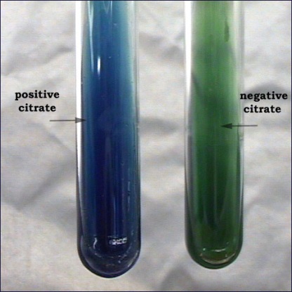
Citrate Test (Simmon's Citrate Slant) Procedure:Streak a loopful of bacteria onto a citrate agar slant, do notstab the butt. Incubate 24 to 48 hours, longer forBacillus species. Incubate with a loose cap. Description:Simmon's citrate agar tests for the ability of an organism to use citrate as its sole source of carbon. This media contains a pH indicator called bromthymol blue. The agar media changes from green to blue at an alkaline pH. Interpretation: After incubation: A positive reaction is indicated by a slant with a Prussian blue color. A negative slant will have no growth of bacteria and will remain green.
İndol, IMViC test grubu içinde yer alan testlerden biridir. IMViC testi; Indol, Metil
kırmızısı, Voges - proskauer ve Sitrat (Citrat) testlerinin ilk harflerinden oluşur. Vi 'deki "i"
küçük harf olarak yazılır ve sadece okuma kolaylığı sağlar. Bu testler, koliform grup
bakterilerin (enterik bakterilerin) ayrımı için kullanılır.
İndol testi; bakterilerin, bir aminoasit olan triptofanı ayrıştırarak indol meydana
getirebilme yeteneğini belirlemek için kullanılır. Aynı zamanda, bakterilerin cins ve tür
ayrımında kullanılır. İndol, bakterilerde bulunan triptofanaz enzimiyle bir aminoasit olan
triptofanı parçalanması ve hidrolize etmesi sonucu oluşan nitrojenli bir bileşiktir ve
aldehidlerle reaksiyona girdiğinde, kırmızı renkli bir ürün oluşturur. Salmonella’da indol
pozitif, Proteus mirabilis’de negatiftir.
Testin yapılış tekniği;
↱ İncelenecek bakteri kolonisinden özeyle alınarak temiz bir tüp içinde
bulunan triptofan bulunan bir sıvı besiyerine ekim yapılır (buyyon veya
peptonlu su)
↱ Tüpler etüvde, 37oC’de 1-5 gün inkübasyona bırakılır.
↱ İnkübasyondan sonra üzerine, tüpün kenarından yavaşça akıtmak
suretiyle 0,5 ml kovaks ayıracı ilave edilir ve tüp çalkalanır. Kovaks
yerine Ehrlich ayıracı da kullanılabilir.
↱ Renk değişimi gözlenir; 1-2 dakika içinde besiyerinin üst kısmında,
parlak kırmızı bir halka oluşması, testin pozitif olduğunu (indol
formasyonunu), sarımsı halka ise testin negatif olduğunu (indol
oluşmadığını) gösterir.
Metil Kırmızısı Testi
Metil kırmızısı testi, besiyerinde bakterilerin glukozu fermente edip organik asit
oluşturarak besiyerinin pH'ını düşürme (4.4'ün altına) esasına dayanır. Bu test,
bakterilerin cins ve türlerinin ayırt edilmesinde kullanılır. Metil kırmızısı testinde;
Escherichia coli’de pozitif, Klebsiella pneumoniae’da negatiftir.
Testin yapılış tekniği;
➤ İncelenecek bakteri kolonisinden öze ile alınarak Glukoz-Fosfat Broth
besiyerine ekim yapılır.
➤ Tüpler etüvde, 37oC’de 1-7 gün inkübasyona bırakılır.
➤ İnkübasyondan sonra üzerine 4-5 damla metil kırmızısı ayıracı damlatılır
ve iyice karıştırılır.
➤ Besiyerinde; kırmızı renk oluşması testin pozitif olduğunu, sarı renk
oluşması ise testin negatif olduğunu gösterir.
➤ Metil kırmızısı ayıracı; pH 4.4 ve altında kırmızı renk, pH 6.0 ve
üzerinde ise sarı renk verir.
Sitrat Testi
Bazı bakteriler karbon-enerji kaynağı olarak sitrat kullanır. Sitrat testi, bakterinin tek
karbon kaynağı olarak sitratı kullanıp kullanmadığını, aynı zamanda bakterinin cins ve
türlerini belirlemek için yapılır. Bakteriler tarafından sitratın metabolizması, citritase veya
citrate demolase adı verilen enzimleriyle gerçekleşir. Klebsiella pneumoniae’da sitrat pozitif,
Escherichia coli’de negatiftir.
Testin yapılış tekniği;
➣ İncelenecek bakteri kolonisinden iğne öze ile alınarak tüpteki Simon’s
Sitrat Yatık Agar besiyerinin yüzeyine ekim yapılır.
➣ Tüpler etüvde, 37oC’de 2-7 gün inkübasyona bırakılır.
➣ İnkübasyon sonunda, orijinal rengi yeşil olan ve indikatör olarak %0,2’lik
Bromo timol mavisi kullanılan besiyerinde, besiyerinin rengi maviye
dönüşür ve pozitif olarak değerlendirilir.
Voges - Proskauer (VP) Testi
Bu test, bakteri türlerini belirlemede kullanılır. Bazı bakteriler, glikozu parçalayarak
nötral bir ürün olan asetil metil karbinol (acetoin) oluşturur. Nöral ürünler
identifikasyonda önemli göreve sahiptir. Asetil metil karbinol, potasyum hidroksid (KOH)
varlığında okside olarak diasetil meydana getirir. Bu ürün ise alfa naftol (kreatin, arginin
veya kreatinin) ile reaksiyona girdiğinde kırmızı renk oluşturur. Klebsiella pneumoniae’da
pozitif, Escherichia coli’de negatiftir.
Testin yapılış tekniği;
⟹ İncelenecek bakteri kolonisinden öze ile alınarak tüpteki Glukoz-Fosfat
Broth besiyerine ekim yapılır.
⟹ Tüpler etüvde, 37oC’de 1-7 gün inkübasyona bırakılır.
⟹ İnkübasyondan sonra üzerine, 1 ml %40’lık KOH, daha sonra 3 ml
%5’lik alfa naftol ilave edilir ve karıştırılır.
⟹ Besiyerinin hava ile temas etmesi için kuvvetli çalkalandıktan sonra 2-5
dakika içinde pembe-kırmızı renk oluşması testin pozitif olduğunu
gösterir.