Microbiology Laboratory Turkey

Mikrobiyoloji Ile Ilgili Tüm Konuların Kısa ve Öz Anlatımları. Microbiology Lab Information.

Bu Blogda Ara(SEARCH)

MicroLab

imvic etiketine sahip kayıtlar gösteriliyor. Tüm kayıtları göster

19 Ekim 2018 Cuma

Ekim 19, 2018

Shigella spp. (TÜRKÇE)

SHIGELLA SPP.

İnsan bağırsağında yaşayan Shigella basilleri, maymunlar dışında sadece insanlarda hastalık meydana getirir. Shigella cinsi içinde Shigella dysenteriae (en ağır seyirli olan), Shigella flexneri, Shigella boydii, Shigella sonnei olmak üzere 4 tür bakteri bulunur.


Morfolojisi

Shigella hareketsiz, sporsuz, kapsülsüz, gram negatif (-) basildir. Hareketsiz olmaları ile Salmonellalardan ayrılır; bakteriyolojik boyalarla kolay boyanır, Shigella flexneri'nin bazı tipleri dışında, Shigella basillerinde fimbria yoktur.


Üreme Özellikleri

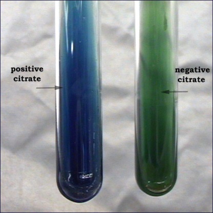
Shigella, aerob veya fakültatif anaerob bakteridir. Optimal üreme ısısı 37oC‟dir; ancak 8-40 oC‟de de üreyebilir. Optimal pH‟sı 7.2-7.4‟dür. Gaita içinde bulunan koliform bakteriler tarafından hızla asitleştirildiği için birkaç saatte ölür. Dış ortam koşullarında oldukça dirençlidir. Isıya, güneş ışığına ve antiseptiklere dirençli değildir. Klorlu sularda harap olur. Adi besiyerinde kolay ürer, buyyonda homojen bulanıklık yapar. Jelozde yuvarlak, hafif kabarık, düzgün yüzeyli, saydam, E.coli kolonilerine benzer S tipi koloni oluşturur. Shigella sonnei kolonileri, diğerlerine göre daha büyüktür. Endo, EMBMacConkey besiyerlerinde renksiz koloni oluşturur. S.sonnei hariç, laktoz negatiftir (-).TSİ besiyerinde asit yapar, gaz oluşturmaz, H2S yapmaz, üre ve sitrat negatiftir. Shigella dysenteriae hariç, diğer Shigellalar mannit (+) tir.

Antijenik Özellikleri
Shigellalarda, O antijeni vardır. Shigella dysenteriae‟nin ekzotoksin ve endotoksini vardır. Bu toksinler, nöro ve enterotoksin (sinir ve bağırsak üzerine etkili) yapısındadır.
Yaptığı Hastalıklar
Shigella dysenteriae basilli, dizanteriye neden olur.


Laboratuvar Teşhisi
➤İnceleme örnekleri: Taze gaita, rektal sürüntü.
➤Teşhis yöntemleri
Mikrobiyolojik örneğin direkt mikroskobik incelemesi 
Taze gaitanın kanlı ve mukuslu kısımlarından hazırlanan preparat, direkt olarak mikroskopta incelenir. Amipli dizanteriden ayırt edebilmek için özellikle Entamoeba histolitica'nın kist ve trofozoit formları ile Giardia, Trichomonas ve Balantidium coli gibi protozoalar araştırılır. Basilli dizanteride gaitada bol lökosit, eritrosit, epitel hücreleri görülür. Mukus bulanık ve kan ortasında ayrı durur. Amipli dizanteride ise az lökosit bulunduğundan mukus daha parlak ve kanla karışıktır.
Rektal sürüntü alınan eküvyonlar stuart saklam besiyerine daldırılırak laboratuvara gönderilir. Temiz bir lam üzerine mukuslu ve kanlı kısımdan örnek alınır, üzerine lamel kapatılır. Lam önce ısıtılır, sonra el değecek kadar soğutularak varsa amiplerin hareketi aktif hale getirilir, preparat mikroskopta incelenir. Entamoeba histolitica ve diğer protozoalar araştırılır.

➤Kültür; taze gaitanın kanlı ve mukuslu kısımlarından Selenit-F, ENDO, EMB, MacConkey, SS, deoksikolat sitratlı agar besiyerlerinden birine tek koloni ekimi yepılır. 24 saat sora üreyen şeffaf, renksiz, küçük koloniler Shigellalar için şüphelidir. Daha sonra bu kolonilerden saf kültür yapılır.
Biyokimyasal testler; lam aglütinasyonu, TSİ, üresitrat, indol, mannit ve hareket testleri yapılır


INSTAGRAM
FACEBOOK 
TWITTER

Ekim 19, 2018

SHIGELLA SPP.

SHIGELLA


Morphology   
➧Small Gram Negative (-), facultatively anaerobic, coliform bacillus
 ➧Non-motile (no H antigen)
 ➧Possess capsule (K antigen) and O antigen
  ➤ K antigen not useful in serologic typing, but can interfere with O antigen determination
  ➤ O antigens: A, B, C, D correspond respectively to the four species
➧Non-lactose fermenting
➧Bile salts resistant: trait useful for selective media.


Taxonomy:
➤ Family Enterobacteriaceae
➤ All Enterobacteriaceae
➧ferment glucose
➧reduce nitrates (NO3 to NO2 or all the way to N2)
➧are oxidase negative


 Four species:
➧Shigella dysenteriae: causes most serious form of bacillary dysentery
➧Shigella flexneri: most common cause of shigellosis in underdeveloped countries
➧Shigella sonnei: most common cause of shigellosis in developed countries
➧Shigella boydii


Clinical Syndromes (Shigellosis):
  Ranges from asymptomatic infection to severe bacillary dysentery.
  Two-stage disease: watery diarrhea changing to dysentery with frequent small stools with blood and mucus, tenesmus, cramps, fever
Early stage:
➧Watery diarrhea attributed to the enterotoxic activity of Shiga toxin
➧Fever attributed to neurotoxic activity of toxin
➧Process involves:
1-Ingestion
2-Noninvasive colonization and cell multiplication
3-Production of the enterotoxin by the pathogenic bacteria in the small intestine;


  Second stage:
➤ Adherence to and tissue invasion of large intestine.
➤ Typical symptoms of dysentery.
➤ Cytotoxic activity of Shiga toxin increases severity.


Epidemiology:
➧ Shigellosis is a major cause of diarrheal disease throughout the developing nations of the world
➧ Major cause of bacillary dysentery (severe second stage form of shigellosis) in pediatric age group (1-10 years old) via fecal-oral transmission
➧ Estimated 15%-20% of pediatric diarrhea in United States
➧ Leading cause of infant diarrhea and mortality (death) in developing countries
➧ Shigella occurs naturally in higher primates, so shigellosis is spread from human to human via the fecal-oral route primarily by contaminated hands (ingestion is portal of entry)
➧ Less frequently, transmission by ingestion of contaminated food or water
➧ Outbreaks usually occur in close communities; Daycare facilities account for a large proportion of cases that occur in the U.S.
➧ Secondary transmission occurs frequently
➧ Low infectious dose (102-104 CFU) with 1-3 day incubation period
➧ Carriage of the organism persists for approximately one month following convalescence


Laboratory Identification:
➧ Closely related to Escherichia
➧ Four distinct species differentiated on basis of serogrouping and biochemical analysis
➧ Stool specimens and rectal swabs should be cultured soon after collection or placed in appropriate transport medium (e.g., Cary-Blair medium)
➧ Readily isolated on selective/differential agar media (e.g., XLD, SS, and brilliant green agar
➧ Lactose nonfermenter


Treatment, Prevention & Control:
➤Dehydration is problem to attend
➤Treat carriers, major source of organisms; Ampicillin and trimethoprim-sulfamethoxazole are effective antibiotics that reduce duration of carriage
➤Antibiotic resistance is a major problem
➤Proper sewage disposal and water chlorination
➤Oral vaccines of Shigella: E.coli hybrids or Shigella mutants offers immunity for six months to one year.


INSTAGRAM
https://www.instagram.com/microbiologylab_turkey/
FACEBOOK 
https://www.facebook.com/mikrobiyolojilab/
TWITTER
https://twitter.com/MicrobioLabTurk


16 Ekim 2018 Salı

Ekim 16, 2018

KLEBSIELLA SPP.

KLEBSIELLA

The genus Klebsiella consists of non-motile, aerobic and facultatively anaerobic, Gram negative rods. At the time of writing, the genus Klebsiella comprises K. pneumoniae subsp. pneumoniae, K. pneumoniae subsp.ozaenae, K. pneumoniae subsp. rhinoscleromatis, K. oxytoca, K. ornithinolytica, K. planticola, and K. terrigena. However, comparison of the sequences of each species shows that the genus is heterogeneous, and may be more reasonably arranged in three clusters. Cluster 1 contains the three subspecies of  K. pneumoniae, cluster 3 contains K. oxytoca and cluster 2 contains the other species (which are notable for growth at 10° C and utilization of L-sorbose as a carbon source). It has been proposed that the genus Klebsiella be divided into two genera and one genogroup, with the name Raoultella being the genus name for those organisms in cluster 2.

For the purpose of conforming to current clinical usage, the clinically important species and subspecies will be referred to as K. pneumoniae, K. ozaenae, K. rhinoscleromatis and K. oxytoca in this chapter.

KLEBSIELLA ON MACCONKEY AGAR

LABORATORY DIAGNOSIS

The members of the genus Klebsiella are Gram negative, nonmotile, facultative anaerobic rods ranging from 0.3 to 1.0 μm in width to 0.6-6.0 μm in length. Most strains grown readily on standard media, although occasionally cysteine requiring urinary isolates of K. pneumoniae are encountered. These strains will appear as pinpoint colonies on routine media, and require supplementation of media with cysteine for more adequate growth. The vast majority of Klebsiella spp. are encapsulated - contrary to popular belief it is probably not capsule which primarily contributes to the mucoid appearance that some Klebsiella strains exhibit.  The Klebsiella which has been linked to the invasive syndrome presenting as liver abscess have a mucoid appearance.

In practice, K. pneumoniae and K. oxytoca are distinguished by indole production by K. oxytoca but not K. pneumoniae. It should be noted however that K. ornitholytica is also an indole producer. The five clinically important species can be distinguished by tests for indole production, ornithine decarboxylase production, he Voges-Proskauer reaction, malonate utilization and o-nitrophenyl-β-D-galactopyranoside (ONPG) production.

KLEBSIELLA ON BLOOD AGAR


Production of plasmid-mediated extended-spectrum beta-lactamases (ESBLs) by Klebsiella spp. has become a major problem. The nature and characteristics of ESBLs are described in greater detail below. Detection of ESBLs in clinical isolates of Klebsiella spp. is problematic since a significant proportion of ESBL producing isolates appear susceptible to third generation cephalosporins or aztreonam. Yet, poor clinical outcomes have been observed when these same antibiotics have been used to treat serious infections due to apparently susceptible ESBL producers. A single surrogate marker for ESBL production, such as ceftazidime resistance, is insufficient for the detection of ESBLs. Virtually all reliable laboratory tests used for detection of ESBLs rely on the change in in vitro activity of oxyimino containing beta-lactams in the presence of a beta-lactamase inhibitor such as clavulanic acid. Examples of ESBL detection methods include the double disk diffusion test, Etest strips containing ceftazidime or cefotaxime with and without clavulanic acid, the Vitek ESBL detection card and the Microscan ESBL plus detection system. Clinical and Laboratory Standards Institute (CLSI) has also developed screening and confirmatory tests for detection of ESBLs. It should be noted that these are standardized for K. pneumoniae and K. oxytoca only.


In some circumstances there is a need to detect ESBL producing Klebsiella spp. from stool or rectal swabs. Examples of such media include Drigalski agar supplemented with cefotaxime 0.5 mg/L , MacConkey agar supplemented with ceftazimide 4 mg/L and nutrient agar supplemented with ceftazimide 2 mg/L, vancomycin 5 mg/L and amphotericin B 1667 mg/L.


INSTAGRAM
FACEBOOK 
TWITTER

4 Ekim 2018 Perşembe

Ekim 04, 2018

TRIPLE SUGAR IRON (TSI AGAR) (ENGLISH)

TRIPLE SUGAR IRON (TSI AGAR)

Triple sugar iron agar (TSI) is a differential medium that contains lactose, sucrose, a small amount of glucose (dextrose), ferrous sulfate, and the pH indicator phenol red.  It is used to differentiate enterics based on the ability to reduce sulfur and ferment carbohydrates. 

As with the phenol red fermentation broths, if an organism can ferment any of the three sugars present in the medium, the medium will turn yellow.  If an organism can only ferment dextrose, the small amount of dextrose in the medium is used by the organism within the first ten hours of incubation. After that time, the reaction that produced acid reverts in the aerobic areas of the slant, and the medium in those areas turns red, indicating alkaline conditions. The anaerobic areas of the slant, such as the butt, will not revert to an alkaline state, and they will remain yellow. This happens with Salmonella and Shigella. 


Procedure:

  1. Touch a well isolated colony with a sterile straight wire.
  2. Inoculate TSI by first stabbing through the centre of the medium to the bottom of the tube and then streak the surface of the slant.
  3. Leave the cap loose and incubate the tube at 35 in ambient air for 18 to 24 hours.
  4. Observe the reaction


INSTAGRAM

FACEBOOK


Ekim 04, 2018

TRIPLE SUGAR IRON (TSI AGAR)

TRIPLE SUGAR IRON (TSI AGAR)

Glikoz, laktoz ve sükroz olarak bakılır. Yatık TSI besiyerine inoküle edilen suş 37°C’ da 24 saat inkübe edilir. İnkübasyon sonunda tüpün dibindeki sarıya dönen renk değişimi glikozun kullanıldığını, yatık yüzeydeki sarıya dönen renk değişimi laktozun kullanıldığını, yatık yüzeyin ucundaki sarıya dönen renk değişimi sukrozun kullanıldığını gösterir. Ayrıca (Norveç üçlü tüp metodunda kullanılan ikinci besiyerine) mannitol’ e geçilerek 37°C’ da 24 saat inkübasyondan sonra sarıya dönen renk değişimi ile mannitol fermantasyonu saptanır.


Testin yapılış tekniği; 
➤ Katı besiyerinde üremiş olan kolonilerden hangi bakteri olduğu tahmin edilenler, TSI agara ekilir. 
➤ Etüvde, 37oC’de 24 saat inkübe edilir.
➤ İnkübasyon sonucunda üreyen kolonilerin gözlenmesi durumuna göre değişen rengin hangi bakteri olup olmadığına karar verilir.


INSTAGRAM

FACEBOOK


1 Ekim 2018 Pazartesi

Ekim 01, 2018

UREASE TEST

UREASE TEST

Objective

To determine the ability of the organism to hydrolyse urea by the action of urease enzyme.


Principle

Urea is a nitrogen-containing compound that is produced during decarboxylation of the amino acid arginine in the urea cycle. Urea is a major organic waste product of protein digestion in most vertebrate and is excreted in urine. Some bacteria have the ability to produce an enzyme urease as part of its metabolism to break down urea. The urease is a hydrolytic enzyme which attacks the carbon and nitrogen bond with the liberation of ammonia and carbon dioxide. It is a useful diagnostic test for identifying bacteria, especially to distinguish members of the genus Proteus from Gram-negative pathogens. Proteus vulgaris is an important and fast producer of urease.

Urease test is performed by growing test organism on urea agar slant or urea broth with phenol red as an indicator with pH6.8. During the incubation period, the organism capable of producing urease enzyme hydrolyse urea and produce ammonia that raises the pH level. As the pH increases, the phenol red changes from yellowish orange as the initial colour of medium to deep pink. Failure of development of a pink colouration due to no ammonia production is evidence of an inability of the organism to produce urease enzyme.

Media

Enzymatic digest of gelatin (1 g), dextrose (1 g), NaCl (5 g), KH2PO4 (2 g), urea (20 g), phenol red (0.012 g), per 1000 mL, pH 6.8.

Procedure

  1. Streak the surface of a urea agar slant with a portion of a well-isolated colony or inoculate test organism on urea broth containing phenol red as the indicator.
  2. Incubate the urea agar slant or urea broth at 37°C for 24-48 hours.
  3. Examine the development of pink colour.
  4. In case of unknown result incubate the tube for 7 days to check for slow urease production.


Results

Positive: Deep pink colouration, Light Orange, Magenta

Negative: No Color change, Yellowish orange colouration


Limitations

  1. Some organisms rapidly split urea (Proteus spp, H. pylori), while others react slowly.
  2. Urea Agar should not be used to determine the quantitative rate of urease activity, as organisms vary in their capability and rate of hydrolysis.
  3. Prolonged incubation may cause an alkaline reaction in the medium and give the false positive result.


Quality Control

Positive: Proteus vulgaris (ATCC13315)

Weak positive: Klebsiella pneumoniae (ATCC13883)

Negative: Escherichia coli (ATCC25922)


INSTAGRAM

FACEBOOK

30 Eylül 2018 Pazar

Eylül 30, 2018

ÜRE TESTİ (UREASE TEST)

ÜRE TESTİ (UREASE TEST)

Bu test, bakterilerin üreyi hidrolize eden üreaz (urease) enzimini belirlemek amacıyla yapılır. Bu bakteriler, üreaz enzim aktiviteleri ile üreyi parçalayarak karbondioksit ve amonyak oluşturur. Oluşan alkali ortamda, besiyerinin pembeye dönmesine neden olur.
Testte Christensen’in üre içeren besiyerleri (üreli buyyon veya agar) kullanılır.

Proteus mirabilis ve Klebsiella üreaz pozitif Escherichia coli negatif bakterilerdir.


Testin yapılış tekniği;

➨ Mikroorganizma kültürlerinden, petri kutusu veya tüpteki Christensen’in üreli agar yüzeyine tekniğine uygun ekim yapılır. 
➨ Etüvde, 37°C’de 1-5 gün inkübe edilir. 
➨ Renk değişmelerine dikkat edilerek her gün kontrol edilir. (Bazı durumlarda renk değişikliği 5-6 saat içinde meydana gelebilir.) 
➨ Kolonilerde veya tüpte pembe rengin meydana gelmesi pozitif reaksiyonu gösterir. 
Negatif durumda renk değişikliği olmaz.  


Üre Testi Pozitif Bakteriler

  1. Proteus spp
  2. Corynebacterium spp
  3. Helicobacter pylori
  4. Brucella spp


INSTAGRAM

FACEBOOK

29 Eylül 2018 Cumartesi

Eylül 29, 2018

IMVIC TEST (Indole Test, Methyl Red Test, Voges Proskauer Test and Citrate Test)

IMVIC TEST
 (Indole Test, Methyl Red Test, Voges Proskauer Test and Citrate Test) 


The IMViC Tests

The IMViC tests are used to differentiate the enterics (Family Enterobacteriaceae). These are the Indole test (tryptone broth), the Methyl Red and Voges-Proskauer tests (MRVP broth) and the Citrate test (Citrate agar slants). For these IMViC tests use the enterics E.coliand Enterobacter.

The significance of these tests is that when testing drinking water for the presence of the sewage indicatorE. coli, one must be able to rule out Enterobacter aerogenes.E. aerogenesis not always associated with sewage, and its presence in water would not necessarily indicate sewage contamination.

Indole Test (Tryptone broth)

Procedure: Inoculate a loopful of bacteria into a tryptone broth. Incubate 48 hours.

Description: Tryptophan hydrolysis -Some bacteria split tryptophan into indole and pyruvic acid using the hydrolase called tryptophanase. Indole can be detected with Kovac's reagent (Indole reagent). This test is very important in differentiating E. coli(indole positive) from some closely related enteric bacteria. It also differentiatesProteus mirabilis(indole negative) from all otherProteusspecies (indole positive). Tryptone broth is used for this test as it contains a large amount of tryptophan.

Interpretation: After incubation: The broth must be turbid. A clear broth indicates that your organism did not grow and cannot be tested. Add a few drops of Indole reagent to the broth culture (tryptone broth). DO NOT SHAKE THE TUBE. A positive result has a red layer at the top. A negative result has a yellow or brown layer.


Methyl Red Test (MRVP broth) 

Procedure: Inoculate a loopful of bacteria into MRVP broth. Incubate 3 to 5 days.

 Description: Mixed acid fermentation -Many gram-negative intestinal bacteria can be     differentiated based on the products produced when they ferment the glucose in MR-VP medium. Escherichia, Salmonella, andProteusferment glucose to produce lactic, acetic, succinic, and formic acids and CO2, H2, and ethanol. The large amounts of acids produced lowers the pH of the medium -Methyl red (a pH indicator) will turn red when added to the medium if the organism was a mixed acid fermenter. Many of these organisms also produce gas.

Interpretation: After incubation: The broth must be turbid. A clear broth indicates that your organism did not grow and cannot be tested. Remove 1 ml of broth and place into a sterile tube before performing the methyl red test ifyou are going to use the same broth for the VP test. Add 3-4 drops of methyl red to the original broth. DO NOT SHAKE THE TUBE. A positive result has a distinct red layer at the top of the broth. A negative result has a yellow layer.


Voges-Proskauer Test

Procedure: Inoculate a loopful of bacteria into MRVP broth. Incubate 3 to 5 days.

Description: Organisms that are negative in the methyl red test may be producing 2, 3 butanediol and ethanol instead of acids. These non-acid products do not lower the pH as much as acids do.Enterobacter, Serratiaand some species of Bacillus produce these substances. There is no satisfactory test for determining production of 2, 3 butanediol. A precursor of 2,3 butanediol called acetoin can be detected with Barritt's reagent.

Interpretation: After incubation: Read the VP test when you have good turbidity. A clear broth indicates that your organism did not grow and cannot be tested. Barritt's reagent A (VP A) contains naphthol and Barritt's B (VP B) contains KOH. Test 1 ml of your culture from the MRVP broth. If you have already conducted the methyl red test, you should have already placed 1 ml of untested broth in a sterile tube. If you haven’t done this, do so now. Add the entire contents of the VP A reagent (15 drops) and 5 drops of the VP B reagent to the 1 ml of your broth culture. SHAKE WELL. This reaction will take a few minutes before you will see a color change. SHAKE the tube every few minutes for best results. With a positive reaction the medium will change to pink or red indicating that acetoin is present. With a negative reaction the broth will not change color or will be copper colored. Wait at least 15 minutes for color to develop before calling the test negative.


Citrate Test (Simmon's Citrate Slant)

Procedure: Streak a loopful of bacteria onto a citrate agar slant, do notstab the butt. Incubate 24 to 48 hours, longer forBacillus species. Incubate with a loose cap.

Description: Simmon's citrate agar tests for the ability of an organism to use citrate as its sole source of carbon. This media contains a pH indicator called bromthymol blue. The agar media changes from green to blue at an alkaline pH.

Interpretation: After incubation: A positive reaction is indicated by a slant with a Prussian blue color. A negative slant will have no growth of bacteria and will remain green.


    IMViC TEST


INSTAGRAM

FACEBOOK

28 Eylül 2018 Cuma

Eylül 28, 2018

IMVIC TEST

IMVIC TEST
(İNDOL, METİLEN KIRMIZISI, SİTRAT, VOGES TEST)


İndol Testi 

İndol, IMViC test grubu içinde yer alan testlerden biridir. IMViC testi; Indol, Metil kırmızısı, Voges - proskauer ve Sitrat (Citrat) testlerinin ilk harflerinden oluşur. Vi 'deki "i" küçük harf olarak yazılır ve sadece okuma kolaylığı sağlar. Bu testler, koliform grup bakterilerin (enterik bakterilerin) ayrımı için kullanılır. 

İndol testi; bakterilerin, bir aminoasit olan triptofanı ayrıştırarak indol meydana getirebilme yeteneğini belirlemek için kullanılır. Aynı zamanda, bakterilerin cins ve tür ayrımında kullanılır. İndol, bakterilerde bulunan triptofanaz enzimiyle bir aminoasit olan triptofanı parçalanması ve hidrolize etmesi sonucu oluşan nitrojenli bir bileşiktir ve aldehidlerle reaksiyona girdiğinde, kırmızı renkli bir ürün oluşturur. Salmonella’da indol pozitif, Proteus mirabilis’de negatiftir.

Testin yapılış tekniği; 
↱ İncelenecek bakteri kolonisinden özeyle alınarak temiz bir tüp içinde bulunan triptofan bulunan bir sıvı besiyerine ekim yapılır (buyyon veya peptonlu su)
↱ Tüpler etüvde, 37oC’de 1-5 gün inkübasyona bırakılır. 
↱ İnkübasyondan sonra üzerine, tüpün kenarından yavaşça akıtmak suretiyle 0,5 ml kovaks ayıracı ilave edilir ve tüp çalkalanır. Kovaks yerine Ehrlich ayıracı da kullanılabilir. 
↱ Renk değişimi gözlenir; 1-2 dakika içinde besiyerinin üst kısmında, parlak kırmızı bir halka oluşması, testin pozitif olduğunu (indol formasyonunu), sarımsı halka ise testin negatif olduğunu (indol oluşmadığını) gösterir.


Metil Kırmızısı Testi 

Metil kırmızısı testi, besiyerinde bakterilerin glukozu fermente edip organik asit oluşturarak besiyerinin pH'ını düşürme (4.4'ün altına) esasına dayanır. Bu test, bakterilerin cins ve türlerinin ayırt edilmesinde kullanılır. Metil kırmızısı testinde; Escherichia coli’de pozitif, Klebsiella pneumoniae’da negatiftir. 

Testin yapılış tekniği; 
➤ İncelenecek bakteri kolonisinden öze ile alınarak Glukoz-Fosfat Broth besiyerine ekim yapılır. 
➤ Tüpler etüvde, 37oC’de 1-7 gün inkübasyona bırakılır. 
➤ İnkübasyondan sonra üzerine 4-5 damla metil kırmızısı ayıracı damlatılır ve iyice karıştırılır. 
➤ Besiyerinde; kırmızı renk oluşması testin pozitif olduğunu, sarı renk oluşması ise testin negatif olduğunu gösterir.
➤ Metil kırmızısı ayıracı; pH 4.4 ve altında kırmızı renk, pH 6.0 ve üzerinde ise sarı renk verir. 


Sitrat Testi 

Bazı bakteriler karbon-enerji kaynağı olarak sitrat kullanır. Sitrat testi, bakterinin tek karbon kaynağı olarak sitratı kullanıp kullanmadığını, aynı zamanda bakterinin cins ve türlerini belirlemek için yapılır. Bakteriler tarafından sitratın metabolizması, citritase veya citrate demolase adı verilen enzimleriyle gerçekleşir. Klebsiella pneumoniae’da sitrat pozitif, Escherichia coli’de negatiftir. 

Testin yapılış tekniği;
➣ İncelenecek bakteri kolonisinden iğne öze ile alınarak tüpteki Simon’s Sitrat Yatık Agar besiyerinin yüzeyine ekim yapılır. 
➣ Tüpler etüvde, 37oC’de 2-7 gün inkübasyona bırakılır. 
➣ İnkübasyon sonunda, orijinal rengi yeşil olan ve indikatör olarak %0,2’lik Bromo timol mavisi kullanılan besiyerinde, besiyerinin rengi maviye dönüşür ve pozitif olarak değerlendirilir.


Voges - Proskauer (VP) Testi 

Bu test, bakteri türlerini belirlemede kullanılır. Bazı bakteriler, glikozu parçalayarak nötral bir ürün olan asetil metil karbinol (acetoin) oluşturur. Nöral ürünler identifikasyonda önemli göreve sahiptir. Asetil metil karbinol, potasyum hidroksid (KOH) varlığında okside olarak diasetil meydana getirir. Bu ürün ise alfa naftol (kreatin, arginin veya kreatinin) ile reaksiyona girdiğinde kırmızı renk oluşturur. Klebsiella pneumoniae’da pozitif, Escherichia coli’de negatiftir. 

Testin yapılış tekniği; 
⟹ İncelenecek bakteri kolonisinden öze ile alınarak tüpteki Glukoz-Fosfat Broth besiyerine ekim yapılır. 
⟹ Tüpler etüvde, 37oC’de 1-7 gün inkübasyona bırakılır. 
⟹ İnkübasyondan sonra üzerine, 1 ml %40’lık KOH, daha sonra 3 ml %5’lik alfa naftol ilave edilir ve karıştırılır. 
⟹ Besiyerinin hava ile temas etmesi için kuvvetli çalkalandıktan sonra 2-5 dakika içinde pembe-kırmızı renk oluşması testin pozitif olduğunu gösterir.


IMViC TEST 

INSTAGRAM

FACEBOOK