Microbiology Laboratory Turkey

Mikrobiyoloji Ile Ilgili Tüm Konuların Kısa ve Öz Anlatımları. Microbiology Lab Information.

Bu Blogda Ara(SEARCH)

MicroLab

microorganisms etiketine sahip kayıtlar gösteriliyor. Tüm kayıtları göster

10 Aralık 2018 Pazartesi

Aralık 10, 2018

Adeno-Rota Virüs Testi

Adeno Rota Virüs Testi

Adenoid dokuda keşfedilen Adenovirüsler; faranjite, farma konjuktival ateşe ve zatürreye sebep olabilir. 5 yaşın altındaki çocuklarda görülen akut solunum hastalığının % 5'inden, bebeklerde görülen zatürrenin % 10'undan sorumludur. Akut gastroenterit olan çocuklarda bakılan gaitanın % 4-14'ünde Adenovirüs tespit edilmiştir.


Rotavirüs, çocuklarda görülen ishale en çok neden olan bir virüstür.  Virüs, ince bağırsağa yerleşerek, çok şiddetli ishal ve kusmayla ortaya çıkar. İleri derecede sıvı kaybı, böbrek yetmezliğine hatta ölümlere bile sebep olur. Özellikle pediatri kliniklerinde yayıldığında kontrol etmek zordur. Erken ve güvenilir tespit Rota virüslerin ileri enfeksiyonlarını önlemede büyük önem taşır.

Amaç 
Gaitada Adenovirüslerin ve/veya Rotavirüslerin kalitatif olarak tespit edilmesidir. 
Prensip 
Bu test, tek basamaklı immunokromotografik yan-akış testidir. Her iki virüse karşı yöneltilmiş monoklonal antikorlar kendi kendilerine Rota virüse özel; kırmızı ya da Adenovirüse özel; mavi lateks partiküllere karşı bağlanır. İki patojene karşı diğer özel antikorlar sıkıca membrana bağlanır. Gaita numunesi ekstrasyon bufer ile karıştırılıp çöktürülür. Temiz sıvı süpernatant test kasedi üzerine alınır. Numune antijene bağlı latex partikülle kasete geçer. Eğer test pozitif ise özel yakalama bandına bağlanır. 

Araç ve Gereçler 
  • Test kaseti 
  • Kullanıma hazır dilüent (Extrasyon bufer) 
  • Tek kullanımlık pipet  
  • Gaita süspansiyonu için test tüpleri 
  • Karıştırıcı 
  • % 0.5 sodyum hipoklorit solüsyonu içeren atık kabı 


Teknik 
  • 1 ml dilüent, test tüpüne konur. 
  • Gaitadan bir miktar alınıp dilüentin içine konur. 
  • Numune iyice homojen hale getirilir. Temiz bir süpernatant oluşana kadar 3 dakika beklenir. 
  • Test kasetleri, oda sıcaklığına getirilir. 
  • Test başlamadan hemen önce test kaseti, poşetinden çıkarılıp düz bir zemin üzerine yerleştirilir. 
  • Pipet ile 4 damla karışım kasetin numune gözüne (S harfi yazan bölüme) damlatılır. 
  • Cihazın numuneyi emmesi beklenir. 


Testin Değerlendirilmesi 
  • Test sonucu, 5 dakika sonra değerlendirilir. 
  • Sonuç penceresinde “C” harfi üzerinde kalan bölümde yer alan tek çizgi yeşil ve “A” harfi üzerinde kalan bölümde yer alan tek çizgi mavi renk Adenovirüs pozitifi gösterir. 
  • Sonuç penceresinde “C” harfi üzerinde kalan bölümde yer alan tek çizgi yeşil ve “R” harfi üzerinde kalan bölümde yer alan tek çizgi kırmızı renk Rotavirüs pozitifi gösterir. 
  • Sonuç penceresinde “C” harfi üzerinde kalan bölümde yer alan tek çizgi yeşil, “A” harfi üzerinde kalan bölümde yer alan tek çizgi mavi ve “R” harfi üzerinde kalan bölümde yer alan tek çizgi kırmızı renk Adenovirüs ve Rotavirüs pozitifi gösterir.
  • Sonuç penceresinde “C” harfi üzerinde kalan bölümde yer alan tek çizgi yeşil, Adenovirüs ve Rotavirüs negatifi gösterir.
  • Test edilen gaita örneğinin fazla olması, sonuç penceresindeki üç bölümde de spesifik bant renklerinin yerine kahverengi rengin oluşmasına neden olur. (Bir anlamı yoktur, daha az miktarda gaita ile test tekrar edilmelidir.)
  • Hiçbir bantda herhangi bir renk görülmez ya da yukarıda tarif edilen, şekillerde gösterilen kombinasyonlardan farklı renkler olursa bir anlam ifade etmez ve değerlendirilmez.
  • Banttaki renk değişimi 10 dakika ya da daha sonra olursa bir anlam ifade etmez ve değerlendirilmez.

Testin Yapılışında Dikkat Edilmesi Gereken Kurallar 
  • Test kaseti 2-30 oC'de saklanabilir. Dondurulmamalıdır. 
  • Test kasetleri ve numune kullanmadan önce oda sıcaklığına getirilmelidir. 
  • Miadı geçen kasetler kullanılmamalıdır. 
  • Buffer solüsyonu sodyum azid içerdiğinden deriye temasından sakınılmalıdır. 
  • Numuneler eldiven giyilerek çalışılmalı ve test bittiği zaman eller yıkanmalıdır. 
  • Test çalışılırken sigara içilmez, herhangi bir şey yenilip içilmez. 
  • Gaita numuneleri hiçbir katkı maddesi olmayan temiz bir kaba konur.


INSTAGRAM

4 Aralık 2018 Salı

Aralık 04, 2018

PROTOZOA

PROTOZOA

Protozoan parasites consist of a single "cell-like unit" which is morphologically and functionally complete and can perform all functions of life. They are made up of a mass of protoplasm differentiated into cytoplasm and nucleoplasm. The cytoplasm consists of an outer layer of hyaline ectoplasm and an inner voluminous granular endoplasm. The ectoplasm functions in protection, locomotion, and ingestion of food, excretion, and respiration. In the cytoplasm there are different vacuoles responsible for storage of food, digestion and excretion of waste products. The nucleus also functions in reproduction and maintaining life. 

The protozoal parasite possesses the property of being transformed from an active (trophozoite) to an inactive stage, losing its power of motility and enclosing itself within a tough wall. The protoplasmic body thus formed is known as a cyst. At this stage the parasite loses its power to grow and multiply. The cyst is the resistant stage of the parasite and is also infective to the human host.

Reproduction – the methods of reproduction or multiplication among the parasitic protozoa are of the following types: 
1. Asexual multiplication: 
(a) Simple binary fission – in this process, after division of all the structures, the individual parasite divides either longitudinally or transversely into two more or less equal parts.  
(b) Multiple fission or schizogony – in this process more than two individuals are produced, e.g. asexual reproduction in Plasmodia. 

 2. Sexual reproduction: 
(a) Conjugation – in this process, a temporary union of two individuals occurs during which time interchange of nuclear material takes place. Later on, the two individuals separate. 
(b) Syngamy – in this process, sexually differentiated cells, called gametes, unite permanently and a complete fusion of the nuclear material takes place. The resulting product is then known as a zygote.

Protozoa are divided into four types classified based on their organs of locomotion. 
These classifications are: amoebas, ciliates, flagellates, and sporozoans. 

3 Aralık 2018 Pazartesi

Aralık 03, 2018

Slayt ile ASO Testi

 Slayt ile ASO Testi

Slâyt üzerinde aglütinasyon yöntemi ile ASO düzeyini ölçmektir.

Araç Gereçler 
  • Reaktif (ASO-Latex (streptolisin O ile kaplı polistiren partikülleri süspansiyonu) 
  • Taze serum numunesi 
  • Pozitif ve negatif kontrol serumları 
  • Tek kullanımlık serum damlatıcı 
  • Slâyt 
  • Cam baget 
  • Rotatör 

Teknik 
  • Tüm reaktifler ve numune, oda sıcaklığına getirilip kullanmadan önce iyice karıştırılır.  
  • Slâyt üzerine bir damla serum damlatılır. 
  • Serum üzerine bir damla ASO-Latex antijeni ekleyip cam bagetle iyice karıştırılır. 
  • Slâyt, iki dakika boyunca hafif dairesel hareketlerle sallanır ve güçlü bir ışık altında aglütinasyon izlenir. 
  • Pozitif ve negatif kontrol serumları da aynı işleme tabi tutulur. 


Testin Değerlendirilmesi
  •  Aglütinasyon görülmesi, pozitif sonucu verir. Pozitif sonuç; numune içerisindeki ASO konsantrasyonunun 200 IU/ml‟ye eşit veya bu orandan daha fazla (normalden yüksek) olduğunu gösterir. 
  • Aglütinasyon görülmemesi,  negatif sonucu verir. Negatif sonuç, numune içerisindeki ASO seviyesinin 200 IU/ml‟den az (normal seviyede) olduğunu gösterir. 


INSTAGRAM

1 Aralık 2018 Cumartesi

Aralık 01, 2018

VDRL TEST

VDRL TEST 

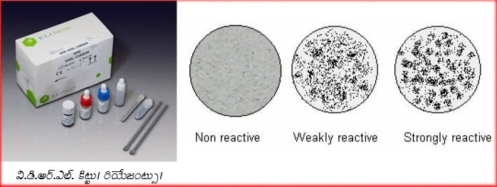
Venereal Disease Research Laboratory (VDRL) Test is a slide flocculation test employed in the diagnosis of syphilis. Since the antigen used in this test is cardiolipin, which is a lipoidal extracted from beef heart, it is not a specific test. This test is also classified as non-specific or non-treponemal or standard test. The antibodies reacting with cardiolipin antibodies have been traditionally (but incorrectly) termed “regain”.


Principle: Patients suffering from syphilis produce antibodies that react with cardiolipin antigen in a slide flocculation test, which are read using a microscope. It is not known if the antibodies that react with cardiolipin are produced against some lipid component of Treponema pallidum or as a result of tissue injury following infection. 

Requirements: Patient’s serum, water bath, freshly prepared cardiolipin antigen, VDRL slide, mechanical rotator, pipettes, hypodermic syringe with unbeveled needle and microscope. Known reactive and non-reactive serum controls are also required. 

VDRL antigen: The cardiolipin antigen is an alcoholic solution composed of 0.03% cardiolipin, 0.21% lecithin and 0.9% cholesterol. The cardiolipin antigen must be freshly constituted each day of test. The working antigen is a buffered saline suspension of cardiolipin. 

VDRL slide: This is a glass slide measuring 2 X 3 inch with 12 concave depressions, each measuring 16 mm in diameter and 1.75 mm deep. 

Procedure: Patients’ serum is inactivated by heating at 56o C for 30 minutes in a water bath to remove non-specific inhibitors (such as complement). The test can be performed both qualitatively and quantitatively. Those tests that are reactive by qualitative test are subjected to quantitative test to determine the antibody titres. 

Qualitative test: 0.05 ml of inactivated serum is taken into one well. 1/60th ml (or 1 drop from 18 gauge needle) of the cardiolipin antigen is then added with the help of a syringe (unbeveled) to the well and rotated at 180 rpm for 4 minutes. Every test must be accompanied with known reactive and non-reactive controls. The slide is then viewed under low power objective of a microscope for flocculation. The reactive and non-reactive controls are looked first to verify the quality of the antigen. Depending on the size the results are graded as weakly reactive (W) or reactive (R). Reactive samples are then subjected to quantitative test. 

Qualitative test: this is performed to determine the antibody titres. The serum is doubly diluted in saline from 1in 2 to 1:256 or more. 0.05 ml of each dilution is taken in the well and 1/60 ml of antigen is added to each dilution and rotated in a rotator. The results are then checked under the microscope. The highest dilution showing flocculation is considered as reactive titre. Sometimes, due to very high level of antibodies in the serum (prozone phenomenon) the qualitative test may be non-reactive. If the clinical findings are strongly suggestive of syphilis, a quantitative test may be directly performed on the serum specimen. 

CSF VDRL: VDRL test may also be performed on CSF samples in the diagnosis of neurosyphilis. Quantitative VDRL is the test of choice on CSF specimens. However, there are some variations in this test. The antigen is diluted in equal volumes with 10% saline, CSF must not be heated (or inactivated), the volume of antigen solution taken is 0.01 ml (or 1 drop from 21 gauge needle) and rotation time is 8 minutes. Rest of the procedure remains same. 

Significance of VDRL test: VDRL test becomes positive 1-2 weeks after appearance of (primary lesion) chancre. The test becomes reactive (50-75%) in the late phase of primary syphilis, becomes highly reactive (100%) in the secondary syphilis and reactivity decreases (75%) thereafter. Treatment in the early stages of infection may completely suppress production of antibodies and result in non-reactive tests. Effective treatment in the primary or secondary stages results in rapid fall in titre and the test may turn non-reactive in few months. Treatment in latent or late syphilis has very little effect on the titre and the titres may persist at low levels for long periods. Since the titre falls with effective treatment, it can be used for assessment of prognosis. VDRL test is more suitable as a screening agent than a diagnostic tool. 

VDRL test is also helpful in the diagnosis of congenital syphilis. Since passively transferred antibodies through placenta may give false reactive test in serum of the infant, a repeat test after a month showing no increase in titre may help rule out congenital syphilis

Since the test employs a non-treponemal antigen, there are many chances of false positive results. False positivity (other than technical) may be due to physiological of pathological conditions. These are called biological false positives (BFP). If the remain positive for less than 6 months it is considered acute and they remain positive for longer than 6 months it is called chronic BFP. The physiological reasons for BFP include pregnancy, menstruation, repeated blood loss, vaccination, severe trauma etc while the reasons for pathological BFP include malaria, infectious mononucleosis, hepatitis, relapsing fever, tropical eosinophilia, lepromatous leprosy, SLE, rheumatoid arthritis etc.

A reactive VDRL test does not necessarily imply that the person is syphilitic. The diagnosis must be made in conjunction with clinical findings. Any reactive VDRL test must be confirmed with a specific or treponemal test such as TPHA, FTA-ABS test. 


INSTAGRAM

30 Kasım 2018 Cuma

Kasım 30, 2018

VDRL Testi

 VDRL (Venereal Disease Research Laboratory) Testi 

  • Venereal = zührevi = cinsel ilişkiyle bulaşan 
  • Disease  = hastalık 
  • Research = İnceleme 
  • Laboratory = laboratuvar 
Treponema pallidum olarak adlandırılan bakterinin neden olduğu sifilis, tedavi edilmediği zaman, vücudun birçok organını etkileyen çok eski çağlardan beri bilinen ve sık rastlanan cinsel yolla bulaşan hastalıklardan biridir. Halk arasında frengi olarak da adlandırılır.  

Treponema pallidum'un organizmaya girişinden yaklaşık 3 hafta sonra şankr denilen ülserel lezyonlar oluşur. Sifilise sebep olan bakteriye karşı organizmanın oluşturduğu antikorlar, (IgG ve IgM) şankr görülmesinden 4-7 gün sonra tespit edilebilir. 

VDRL'nin lamda kalitatif ve kantitatif analizi yapılır, otoanalizörde çalışılır. Günümüzde ise mikrobiyoloji laboratuvarında ve kan merkezlerinde kaset testi ile yapılır. 


INSTAGRAM

29 Kasım 2018 Perşembe

Kasım 29, 2018

TRICHURIS TRICHIURA

TRICHURIS TRICHIURA 
(WHIP WORM) 

The worm is divided into a thin whip-like anterior part measuring 3/5 of the worm and a thick fleshy posterior part of 2/5 the length. 
Male: The male measures 3-4.5 cm in length. Its posterior end is coiled and possesses a single cubicle. 
Female: The female measures 4-5 cm in length. Its posterior end is straight 

Infective stage and mode of infection 
Infection is by ingestion of eggs containing larvae with contaminated raw vegetables. 

Life Cycle: 
 Ingested eggs hatch in the small intestine and the larvae migrate to the large intestine to become adult. After mating, the female lays immature eggs, which pass with the stool to the soil and mature in 2 weeks.  


Symptoms  
 The patient complains of dysentery (blood and mucus in stool together with tenesmus). Rectal prolapse is also possible. 

Diagnosis 
 Finding of characteristic eggs. The egg of trichuris is barrel-shaped, 50x25 microns. The shell is thick with a one mucoid plug at each pole.


INSTAGRAM